A research group led by Professor Shintaro Nakao of the Department of Ophthalmology, Graduate School of Medicine, Juntendo University, conducted a comparative study (First-in-Human) using the intraocular endoscope holding robot "OQrimo"*¹ to observe the peripheral retina without performing "scleral depression"*², which was conventionally essential in vitreoretinal surgery. In conventional surgery, scleral depression was necessary to observe the peripheral retina, which posed a challenge due to the pain and other burdens on patients. In this study, 16 patients were divided into a robot group and a conventional method group for comparison. The results showed that the degree of postoperative inflammation was comparable to the conventional method, and no intraoperative complications occurred, confirming its safety. This achievement not only contributes to the establishment of a new surgical method that reduces the physical burden on patients but also significantly promotes the widespread adoption and development of robotic surgery in the ophthalmology field. This paper was published online in 'Ophthalmology Science' on February 5, 2026.
Highlights of This Research Achievement
● Conducted a comparative study on the usefulness and safety of peripheral retinal observation without scleral depression using the intraocular endoscope holding robot "OQrimo" versus the conventional method.
● The robot group successfully observed the peripheral retina without compression, confirming high safety comparable to the conventional method in evaluating postoperative inflammation, etc.
● Expected to reduce the physical burden on patients and contribute to the future development of ophthalmic robotic surgery.
Background
Observation of the peripheral retina is indispensable for treating lesions and preventing complications in vitreoretinal surgery performed for conditions such as retinal detachment, which can lead to blindness. Traditionally, this observation has involved a technique called "scleral depression," where the white of the eye is pressed from the outside of the eyeball. This technique causes severe pain and discomfort to patients and can lead to postoperative inflammation. Furthermore, it occupies one of the surgeon's hands, limiting delicate operations that require both hands. In recent years, as robotic surgery has become widespread in the medical field, we have been involved in the development of the intraocular endoscope holding robot "OQrimo." The purpose of this study was to verify the safety and usefulness of a new ophthalmic robot-assisted surgery that observes the peripheral retina without performing scleral depression, which is a physical burden on patients, by using OQrimo.
Content
This study targeted 16 patients (16 eyes) scheduled for 25-gauge vitrectomy for epiretinal membrane*³ and simultaneous cataract surgery. Patients were divided into a "robot group" (8 eyes), where peripheral retinal observation was performed without scleral depression using the intraocular endoscope holding robot "OQrimo," and a "conventional group" (8 eyes), where the conventional scleral depression method was used, to compare effectiveness and safety. As a result, in the robot group, peripheral retinal observation was successfully performed without scleral depression in 87.5% (7/8 eyes) of cases. The range of peripheral retinal observation by the endoscope held by the robot reached an average of 9.29 (±2.13) hours on a clock face (12 hours), demonstrating that a wide range of observation is possible. Regarding safety, although the operation time in the robot group was longer than in the conventional group, no significant difference was observed between the two groups in the degree of intraocular inflammation (anterior chamber flare value) at 3 hours, 24 hours, and 1 week postoperatively, or in the patient's pain score on postoperative day 1. Furthermore, wound healing was not problematic in the robot group, and no serious surgical complications were observed in either group. These results demonstrate that peripheral retinal observation without scleral depression using an intraocular endoscope holding robot can be performed with high safety comparable to the conventional method. This achievement is significant not only for eliminating painful scleral depression and reducing the physical burden on patients but also for greatly promoting the social implementation of robot-assisted surgery in the ophthalmic field in the future.
Future Development
This time, the research group demonstrated that by using the intraocular endoscope holding robot "OQrimo," the peripheral retina can be safely observed without scleral depression, which has been a physical burden on patients. Although some blind spots remain in the observation with the current robot-held endoscope, it is expected that more comprehensive peripheral observation will be possible in the future with the development of endoscopes with wider fields of view and curved endoscopes. This method allows the operating surgeon to continue using both hands in ophthalmic surgery, which is expected to be applied to current complex vitreoretinal surgeries and to contribute to the development of ophthalmic robotic surgery. We will continue to promote research aimed at further improving the efficiency and social implementation of this system, with the goal of providing less invasive and less physically burdensome safe ophthalmic medical care to more patients.
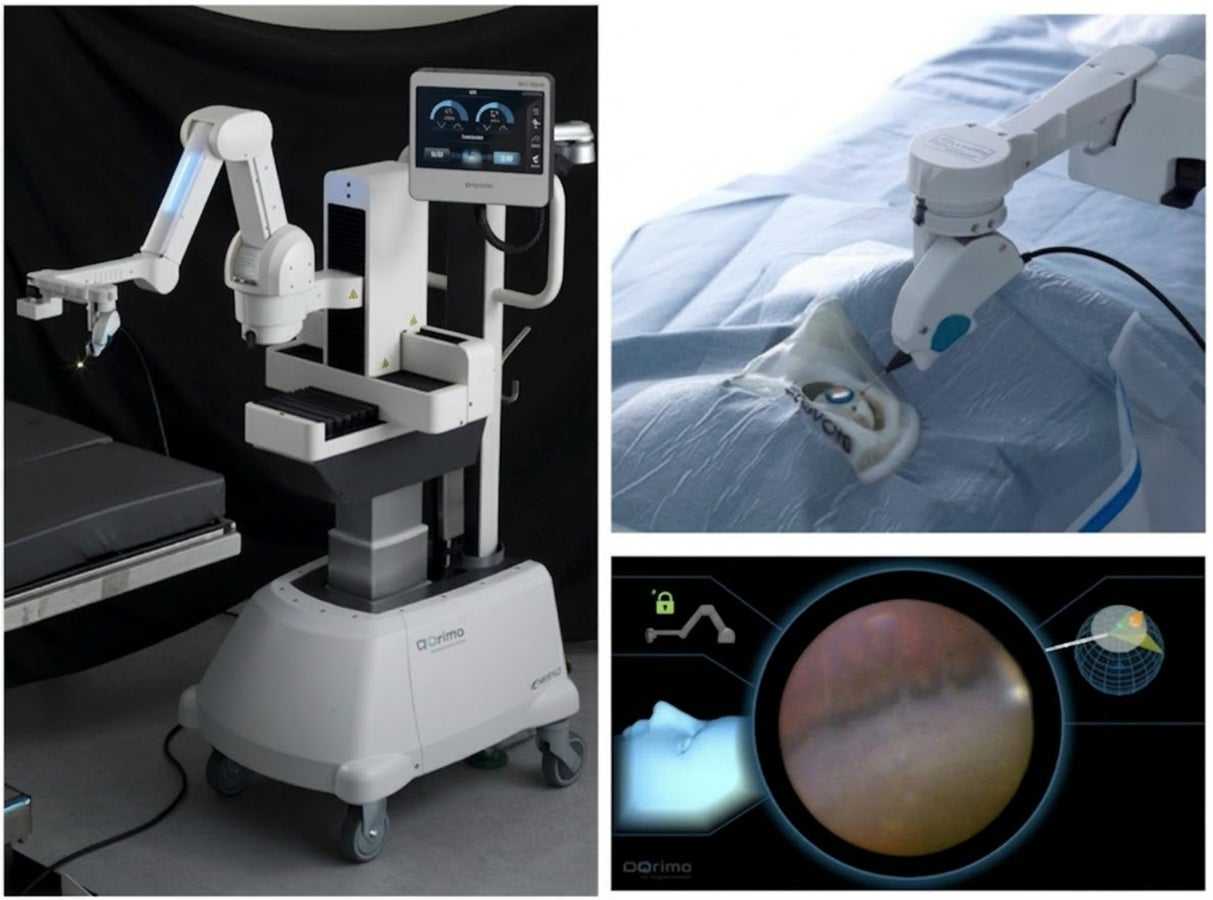
Figure 1: Peripheral retinal observation using the intraocular endoscope holding robot "OQrimo"
(Left) Exterior view of OQrimo. (Top right) Intraocular endoscope held by OQrimo inserted into an eye model. (Bottom right) Actual observation image of the peripheral retina by the endoscope held by OQrimo.
Glossary
*1 Intraocular endoscope holding robot "OQrimo": A medical robot that holds an intraocular endoscope on behalf of the surgeon and allows for fine position adjustments via pedal operation, etc. It enables the surgeon to perform precise surgical operations with both hands while observing areas that can only be seen with an endoscope.
*2 Scleral depression: A technique in vitreoretinal surgery where the eyeball is pressed from the outside (white of the eye) with a specialized instrument to observe the peripheral part inside the eyeball. It often causes pain and discomfort to patients.
*3 Epiretinal membrane: A disease where a fibrous membrane forms on the surface of the macula, the central part of the retina responsible for vision. It causes symptoms such as decreased vision and distorted vision (metamorphopsia).
Researcher's Comment
This research is a significant first step demonstrating the great potential of robot-assisted technology in ophthalmic surgery. In conventional vitreoretinal surgery, a painful procedure was necessary to examine every corner of the retina, and surgeons were limited by having one hand occupied. By using OQrimo, these challenges can be overcome, and it opens the way for ultra-high precision treatments that may exceed the limits of human dexterity in the future. We will continue to advance technology development and evidence building that meets the needs of clinical practice, aiming to contribute to the advancement of ophthalmic medical care.
Original Paper
This research was published online in "Ophthalmology Science" on February 5, 2026.
Title: Robot-Assisted Peripheral Retinal Visualization During Vitrectomy Without Scleral Depression: A First-in-Human Comparative Study
Authors: Kenta Ashikaga1,2, Yoshihito Sakanishi1,2, Toshiaki Hirakata1,2, Shutaro Yamamoto1, Ayumi Usui-Ouchi1,2, Koh-Hei Sonoda3, Shintaro Nakao1,2
Author Affiliations: 1) Department of Ophthalmology, Graduate School of Medicine, Juntendo University, 2) Juntendo He's Advanced Ophthalmic Technology Research Laboratory, 3) Department of Ophthalmology, Graduate School of Medical Sciences, Kyushu University
DOI: 10.1016/j.xops.2026.101105
We express our sincere gratitude to everyone who cooperated in the implementation of this research.
FACT BOX
- Source: PR TIMES
- Category: News